Care of the Horses Hoof, an Overview
by Robert N. Oglesby DVM
Introduction
Introduction
»
Hoof Anatomy and Physiology
»
Nutrition and Health Care
»
Environment: Keep Them Clean and Dry
»
Keep them Trimmed
»
Shoes?
»
More Info & Discussions
You would think that this most important part of the equine anatomy would be the subject of huge amounts of research and investigation. Until very recently however, this was not true. The result is that many of our commonly accepted precepts are at best untested and, as good scientific information is uncovered, some have been shown to be untrue. Good hoof care, though the subject of speculation since the time man first hitched a horse to a load, is still poorly understood. Now don't take this the wrong way: experience has led us to a workable methodology that results in the majority of horses being able to perform their intended work with few hoof problems. It is also true that there are still many chronic hoof problems that defy attempts at correction and sometimes explanation. Some of the known and conjectured problems with poor quality horn:
-
genetics
-
nutrition
-
past trimming and shoeing experiences
-
environment
-
injury
This article provides an overview of the anatomy and principles of hoof care and trimming for horses and other equines. Links are provided to more detailed articles on various subjects pertaining to the care and diseases of horse hooves. Summaries of scientific reports are at the end of this article.
Hoof Anatomy and Physiology
Introduction
»
Hoof Anatomy and Physiology
»
Nutrition and Health Care
»
Environment: Keep Them Clean and Dry
»
Keep them Trimmed
»
Shoes?
»
More Info & Discussions
|
To properly understand the hoof, you have to know its parts and how it grows. The illustration to the right gives you a general idea of the superficial structures around the foot and how the foot gets its blood and nerves. Also the silhouette of the underlying bone of the hoof, the coffin bone, is shown. Important to note is that the majority of the wall is formed at the top on the hoof and grows down, taking somewhere between eight and 12 months to grow out. This is important to realize because any damage introduced into the horn will persist until it grows out. The tissue that actually forms the horn of the foot is called the corium. The coronet, wall, and sole all have horn producing corium tissue. The corium also serves to bind the horn to the living tissue.
|
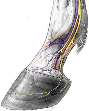
-
nerves = yellow
-
veins = violet
-
arteries = red
-
ligaments and tendons = orange
|
|
Taking a closer look at the top of the wall notice the bulge of the coronary corium that is continuous with the wall corium. The majority of the wall is made here by groups of cells spinning out protein tubules and crosslinking them with adjacent groups of cells. The crosslinks are methionine molecules, an essential amino acid in protein. If you look closely at the diagram, you can see the tubules in the diagram and in your horse's hooves. The wall is pushed distally toward the ground, with the leaves of the insensitive laminae sliding between the sensitive laminae. Looking at the diagram you can see the interdigitation on the insensitive and sensitive laminae. The leaves of the insensitive laminae can be seen growing out the bottom of the foot as the white line of the sole, just behind the wall.
|
|
|
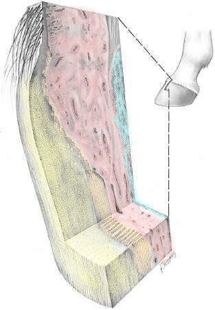
With the horn of the foot (wall, sole, frog) removed you can see the structure of the corium. The corium is the horn producing tissue of the foot and also acts to attach the horn to the underlying bone and tissues. Even though most of the wall is formed at the coronet band, the wall corium adds horn to the wall as it grows down, thickening the wall as it travels distally.
|
 Starting from left to right at the bottom of the image:
-
Wall Horn
-
Laminae: with the interdigitations of the insensitive on the left and sensitive laminae on the right
-
Wall or Lamellar Corium: a very vascular tissue that forms the sensitive laminae and attaches the wall to the bone
-
Periosteum and bone: the thin sliver on the far right of the image
|
|
Here again we can see the relation of the wall and sole to the bone of the foot. Also, important to note are the position on the navicular bone and insertion of the flexor tendon.
|
-
deep digital flexor tendon = yellow
-
navicular bone = blue
-
impar ligament = orange
-
intersection of ligament and tendon = red
|
|
The blood supply to the foot runs down the sides and just slightly to the rear of the pasterns, just in front of the flexor tendons on each side. Recent research has shown that there is a pump system in the back of the foot that works in reverse to what we used to think: Blood is drawn into the foot during weight bearing and pushed out when weight is taken off the foot!
|
|
Let's review the sole anatomy. This is an illustration of a front foot. The hind foot is usually not so round. Of course feet vary in their shape but this would be an ideal. Starting at the outside of the foot:
- Wall.........yellow
- White line...thin line just to the inside of the wall that goes all the way from one heel to the other
- Bars.........blue
- Frog.........the large triangular structure between the bars
- Sulcus of the Frog....red
|
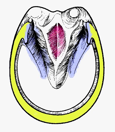 Notice
-
the wide open sulcus and bars
-
that the widest part of the frog lies adjacent to the end of the solar surface of the wall at the heel
-
the relatively short distance from the tip of the frog to the wall at the apex of the toe
|
Nutrition and Health Care
Introduction
»
Hoof Anatomy and Physiology
»
Nutrition and Health Care
»
Environment: Keep Them Clean and Dry
»
Keep them Trimmed
»
Shoes?
»
More Info & Discussions
You have just read the beginning of this article. To access the unabridged article
become a Member of Horseadvice.com! Your membership gets you instant access to this and over 600 articles on our site. Other benefits of your membership include participation in our discussion boards and access to our one button PubMed search tool for each topic.
Horseadvice can teach you to be a more knowledgeable horse owner which leads to a healthier horse and save you money. Really, we guarantee it.
Come Join Us!
 .
.