Correcting Long Toe,
Low Heel Foot Conformation in Horses
by Robert N. Oglesby DVM
Introduction
Introduction
»
Biomechanics
»
Evaluating Breakover
»
Evaluating Heels
»
Corrective Trimming
»
Example
»
More Info & Discussions
Navicular disease or syndrome is a common problem in horses. With the recognition that many structures may be involved with this problem it in now more accurately referred to as "chronic heel pain" or "chronic palmar foot pain". No matter what you call it these horses typically have a slow onset bilateral lameness that improves with posterior digital nerve block
...more. Often there is not a definitive diagnosis of the cause of the pain but many of these horses suffer from abnormal hoof conformation with long toes and underrun heels with a broken hoof pastern axis, more. This conformation is likely to contribute to increased stress of the palmar structures of the foot and therefore predispose to lameness or increase the pain of preexisting lameness.
Trimming these feet to return to a more normal conformation and to ease the breakover of the foot is integral to making these horses as comfortable as possible. And there are other conditions that may be more comfortable with improved foot conformation and easing breakover and would include degenerative joint disease of the lower joints, digital flexor tendonitis, and suspensory disease.
The chronic nature of the poor foot conformation often makes achieving a normal foot difficult with simple trimming and/or shoeing. This article uses case studies to address the common abnormal conformation and ways to assess and trim to improve foot function in conditions where improved foot conformation will help.
Biomechanical Principles of Foot Conformation
Introduction
»
Biomechanics
»
Evaluating Breakover
»
Evaluating Heels
»
Corrective Trimming
»
Example
»
More Info & Discussions
It may help to understand the principles of how conformation effect the biomechanical workings of the horses foot to consider the following thought experiment. Take two pairs of 1 inch thick boards that are as wide as your foot. One pair is one half the length of your foot the other pair twice the length of your foot. One pair at a time strap them on to the bottom of your feet and walk around. Let's start with the short boards. As you move your weight forward in preparation of your first step, the front of your foot rocks down just before your foot would have normally left the ground. This positions the foot in a manner that is roughly that of the foot leaving the ground, aiding in the breakover, that moment your foot first leaves the ground. Now strap the long boards to your feet and repeat the first step. You instantly realize it is much harder to walk requiring greater effort to breakover your foot and overcome the extra length of your foot with the board attached. You can feel the extra pull in the flexors in the back of your legs.
Examine the image to the right and picture the foot is just about to breakover. Forward motion and breakover occur as the flexors pull on the back of the foot. The arrows show some of the areas that are stressed as the flexor tendon is pulled by the more proximal flexor muscles.
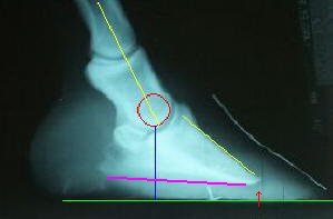
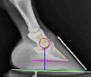
Examine the image and the radiographs. Start with the red circles these represent the center of rotation of the coffin joint as the foot breaks over. The edge of the circle is congruent with the coffin joint. The green line is the length of the sole from the heel to the toe. By dropping a line, here in blue, from the center of rotation to the line that marks the sole length you cut the line and from where it intersects the green line you can get two important pieces of information. The first is the anterior (front) segment of the green line shows you the moment arm of the lever that the muscoskeletal system has to overcome to breakover the foot as the horse moves forward. The longer this line is the more effort required. The posterior segment is proportional to the amount of force placed on the heels when the horse bears weight. The smaller the area the more pressure placed on the heels during weight bearing. Experience suggests that the closer the blue comes to evenly dividing the green line the healthier the foot.
The radiograph on the top shows a poorly conformed hoof with a very long toe and broken hoof-pastern axis (yellow lines are not parallel). Usually hooves like this also have run under heels, not a feature of this foot. Note where the blue line, the projection of the center of rotation onto the sole, intersects the green line, which is the length of the sole. See how much of the green line, as a percentage of the total green line, projects in front of this intersection? This is the board that stick out in front of your foot. Also note how thin the sole is a common finding in horses with this conformation. One last feature of this radiograph is the deformation of the tip of the coffin bone (red arrow). This indicates excessive force being placed on the foot at this point, a result of the thin sole and long toe. It would be surprising if there was not pain originating in this area also.
The radiograph underneath, though not ideal, is much closer to a balanced foot. The yellow lines are near parallel, the blue line intersects the green line closer to the middle, and there is good sole depth. However the solar angle of the coffin bone (the angle the purple line makes with the ground) is quite flat. Normal feet usually have an angle between 5 and 10 degrees. The radiograph of the long toed foot has a better angle but still not as steep as would be considered normal.
Long toes and low heels increase the stress on many of the palmar structures of the foot and leg, particularly during movement. The conformation also may cause degenerative changes in the supporting tissues of the foot including a thin sole, broken down heels and buttresses, DDF tendonitis, desmitis (strain on the ligaments) of the ligaments that support the navicular bone and coffin joint.
Evaluating Toe Length and Breakover
Introduction
»
Biomechanics
»
Evaluating Breakover
»
Evaluating Heels
»
Corrective Trimming
»
Example
»
More Info & Discussions
To read more on this topic become a member of
Horseadvice.com! Your membership gets you instant access to this and over 600 equine articles on our site. Other benefits of your membership include participation in our discussion boards and access to our one button PubMed search tool for each topic.
Horseadvice.com educates you to be a more knowledgeable horse owner which leads to healthier horses and save you money, we guarantee it. Come Join Us!
 .
.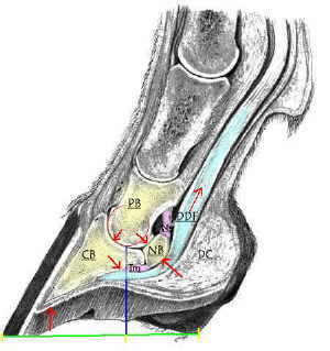 CB: coffin bone; NB: navicular bone; PB lower pastern bone; DDF: deep digital flexor tendon; Im: impar ligament; NS: navicular suspensory ligament;