|
|
Discussion on Dark fluid filled bumps
|
| Author |
Message |
New Member:
mcgover
|
Posted on Tuesday, May 13, 2008 - 12:41 am:
My mare has a couple of these "bumps" on her neck and one on the inside of her leg. I first noticed them last summer and thought maybe it was from an insect bite. I asked my vet when she came out for spring vaccinations but she was unsure what they were. They are raised and fluid filled and about the size of a M & M. She has had them for a year now but they have not changed. Just wondered if anyone knew what they were and how to treat. 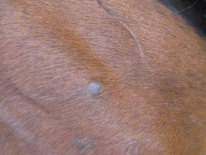
|
Moderator:
DrO
|
Posted on Tuesday, May 13, 2008 - 7:27 am:
Hello Laura,
Though there are lots of possibilities from the outward appearance but the presence of "a dark fluid" on the inside makes these an oddity unless the fluid is midnight black. Occasionally melanomas will fill with a thickish fluid that is a very black. Your veterinarian needs to take a sample of the fluid for analysis, as it's nature may be a key to what the bump is.
DrO
|
New Member:
mcgover
|
Posted on Friday, May 30, 2008 - 4:11 pm:
Hi Dr O,
Just a follow up on the black bumps:
My vet did a fine needle aspiration of the largest bump and the results came back as follows:
Three films evaluated. One film consists of cellular debris and crystalline material only. The other two films are similar with a few small, densely cellular areas containing heterogenous lymphocytes in sheets. In these areas, small lymphocytes predominate and there are occasional plasma cells, neutrophils and intermediate and large lymphocytes. One very small area has a higher proportion of intermediate forms and may represent a follicle. Overall, the findings are consistent with lymphoid hyperplasia or reactive lymphoid tissue. The crystalline material may be artifact or if intralesional, may represent calcification. If this is not consistent with the clinical picture, biopsy with histopathology may be warranted.
Interpretations: Lymphoid Hyperplasia
When I checked lymphoid hyperplasia in horses it is usually dealing with the airway? Do you know of any skin types?
I have noticed the one bump that was aspirated seems to not be as fluid filled. When the vet did the aspiration I was surprised to not see fluid come out but maybe that was because it was a small needle?? There was a little speck of blood where the needle went in but no black fluid.
The vet & I did chart the size and location of the other bumps to see if they change.
The bumps are not bothering my horse.
Do you recommend any type of treatment?
Thanks,
Laura
|
Moderator:
DrO
|
Posted on Saturday, May 31, 2008 - 4:52 pm:
If I were treating a horse with what appeared to be be bumps from a non-infectious immune reaction I would inject each one with 1/4 cc methyl-prednisolone (Depo to most vets). Alternatively a steroid cream twice daily could be used. These need to be discussed with your veterinarian however.
DrO
|
|
|
